|
The following sections are provided as information for patients.
The external ear
This is merely the pinna and auditory meatus.
Diseases of the external ear
There is always a danger of forcing the foreign bodies through into the ear canal where it may damage the drum and the middle ear. Extraction of foreign bodies should therefore be done by skilled ENT personnel.
Infection
Infection of the ear canal could be in the form of a boil which is very painful or generalised infection which could be bacterial, fungal or viral. Reactive inflammation in the form of eczema and dermatitis could also give symptoms of itching, discomfort and ear blockage. In patients for example with diabetes or elderly patients with low immunity the infection can spread deep into the tissues and in the bone.
Treatment of it should consist of thorough aural cleaning with microsuction, ear drops and sometimes antibiotics are necessary.
Otitis Media
Acute otitis media is infection and inflammation behind the eardrum which will lead to pain, hearing loss and sometimes perforation of the eardrum and a discharge through it may develop. In the majority the inflammation resolves and the tympanic membrane heals without any sequelae. However in a small proportion of patients complications develop and there is loss of hearing.
Other problems are :
- Chronic middle ear effusion or glue ear.
- Scarring of the tympanic membrane and around the middle ear ossicles.
- The infection may become persistent and chronic with a non-healing perforation of the tympanic membrane.
- An infection may progress to mastoiditis.
Otitis media with effusion or glue ear
Accumulation of non-purulent fluid in the middle ear behind the tympanic membrane is common in children aged between 2 and 6 years. The most common causes is low grade inflammation with partial blockage and dysfunction of the Eustachian tube and interference with the free flow of air in and out of the middle ear. Adenoids behind the nose could partly be responsible for Eustachian tube blockage and dysfunction.
The symptoms are :
- Impaired hearing
- Delay in hearing to speak
- Acquiring vocabulary and other language difficulties
- Inattentiveness
- Recurrent earache
Management
In many instances reassessment after 3 months is advisable because the majority of the fluid will resolve spontaneously. Unresolved middle ear effusion or glue with hearing loss requires myringotomy (making a small incision in the eardrum) sucking out the fluid through it and inserting a ventilation tube called a grommet. The ventilation tube or grommet remains in the tympanic membrane for about 12 months helping to reduce to normal the mucous producing mucosa in the middle ear and then it spontaneously extrudes from the tympanic membrane.
Middle ear effusion may also occur in adults and needs thorough investigation in case there is anything blocking the tube where it opens behind the nose.
Chronic suppurative otitis media
The discharge through a perforation in the tympanic membrane may be treated with drops and then the perforation could be grafted.
Once the infection spreads into the bone of the mastoid then drops may not help especially if cholesteatoma develops which is a mass of skin tissue adjacent to the eardrum. This requires an operation of mastoidectomy. Once the middle ear bones and the tympanic membrane is destroyed by the infection it may require reconstruction by a tympanoplasty. For reconstruction of the ossicles a prosthesis is used.
Otitis media can give complications such as mastoiditis, infection may spread deep in the inner ear and cause labyrinthitis and dizziness and the infection can also affect the nearby passing nerve of the face and cause a facial palsy. If infection spreads outside the bones of the ear it can cause a brain abscess and meningitis.
The middle ear
The tympanic membrane separates the external from the middle ear. The middle ear cavity communicates with the mastoid air cells. The bony roof of the middle ear separates it from the covering of the brain. The middle ear is in contact with the external atmosphere through the eustachian tube which opens into the space behind the nose and contains three articulating ossicles – malleus, incus and stapes which are supported by ligaments. The ossicilation of the tympanic membrane and a chain of small bones allows the transmission of sound pressure and movement of fluid in the inner ear.
The Inner Ear
There are three parts of the inner ear:
- Anteriorly the cochlea for hearing.
- The middle part, the vestibule which is concerned with static balance and linear acceleration.
- Posteriorly three semicircular canals of balance in different planes which are concerned with angular acceleration. The hearing and balance nerve (VIIIth) joins and travels through the internal auditory meatus to the brain. The facial nerve (VIIth) also travels through the internal auditory canal and traverses the middle ear cavity to emerge through the opening in the base of the mastoid and is responsible for movement of the face.
Balance
Movement and acceleration in any of the three planes of the canals of balance of the inner ear and in the vestibule stimulate receptors which are imbedded in gelatinous material. Changes in linear or angular acceleration cause movement of the sensory cells and that triggers the action potential of the vestibular nerve which travels into the brain. All the input from the inner ear, the eyes and the sensory receptors of the joints and tendons relay to the brain. Central processing takes place and responses return to the muscles to maintain posture and eye position. An alteration in vestibular response on one side as opposed to the other result in imbalance in central response which affects control of the eyes causing them to osscilate/vestibular nystagmus.
Common symptoms:
- Hearing loss;
- Aural discharge;
- Otalgia, or pain in the ear;
- Tinnitus, or variable noises in the ear;
- Vertigo.
When otalgia is a presenting symptom and no local disease is found in the ear, referred pain is possible from a distant area shared by supply of the same nerve and the usual causes are:
- Dental disease;
- Temporamandibular joint dysfunction;
- Maxillary sinusitis;
- Inflammatory and malignant lesions of the throat, posterior part of the tongue and voice box;
- Conditions in the back of the neck and cervical spine.
Tinnitus
The subjective perception of tinnitus which is characterised by a rushing, ringing, hissing sounds of various intensity in the ear or head may be associated with dysfunction in the inner ear with various lesions along the pathway in the brain. Rhythmic pulsatile tinnitus is suggestive of vascular lesions such as:
- Arterial venous malformations;
- Arterial aneurysm;
- Glomus tumours in the middle ear;
- Sound transmission from major vessels in the neck.
Crackling sounds can be associated with dysfunction of the Eustachian tube and rhythmic contractions of the muscles attached to it.
Vertigo
Vertigo associated with peripheral inner ear is most commonly rotatory, but can be experienced as swaying or tilting of either the patient or surroundings. Movement and positional changes tend to make vertigo worse. Central vestibular lesions tend in the brain to produce less intense vertigo, positional changes have less effect and the patient may experience disturbance of gait and other neurological symptoms and signs.
- Labyrinthine nystagmus of the inner ear is always horizontal rotatory or horizontal.
- Vertical nystagmus and multiple other forms occur only in central vestibular lesions in the brain.
- Benign positional vertigo and nystagmus when the defect is thought to be due to a floating otolith particle in one of the canals of balance, occurs with a short latency lasts a few seconds and fatigues on repetition. It can be treated with the Epley vestibular repositioning manoeuvre.
Vestibular tests such as electronystagmography can record the velocity of nystagmus and assess by graphic recording. A caloric test is done to stimulate the balance organ by cooling or heating the labyrinth by flow of air or water in the external ear canal. In vestibular hypofunction these may be reduced or absent.
Hearing
Micromechanical ossicilations of the eardrum and the ossicular chain result in movement of liquid in the inner ear (cochlea) and motion of the basilar membrane with sensory hair cells. The movement of the receptor hair cells generate electric impulses in the cells and bioelectrical events then follow in the auditory nerve.
Hearing Loss
There are three types of hearing loss:
- Conductive loss from lesion in the external auditory canal and the middle ear like ear perforations or disruption of the middle ear ossicles, fluid behind the eardrum or infection.
- Sensorineural hearing loss from cochlea and cochlear nerve and lesions of the auditory pathway in the brain.
- Mixed when both types of hearing loss are present.
Pure tone audiometry can record the hearing threshold by measuring decibels of hearing levels and at each frequency between 250 and 8000 Hz.
Impedence audiometry for the middle ear compliance (i.e. how much of the applied sound energy is reflected from the tympanic membrane) and middle ear pressures (the difference if any between pressure externally and in the middle ear cavity) can be measured with an instrument applied to the ear canal. A very low compliance could be the result of fluid behind the eardrum. Lower or negative pressure within the middle ear than without indicates insufficiency of the eustachian tube and underventilation of the middle ear.
Electric response audiometry to sound stimuli can be recorded from the cochlea, brain stem and cortex and auditory evoked potentials ? can be recorded from the scalp. This has become useful as an objective test in children and in patients in whom reliability of subjective audiometric testing is doubtful. It also has wide ranging application in neuro-otological diagnosis.
Diseases of the inner ear
Otosclerosis
Otosclerosis is a localised diseased of bone which is inherited. Spongy bone forms and if this is in the area of the stapes ossicle there may be fixation of it and stop it moving hence causing conductive deafness. There is a strong family history and both ears are affected in 90% of patients. The tympanic membrane is normal and mobile in the presence of hearing loss on an audiogram.
Management
If the patient does not wish to have a hearing aid then a stapedectomy is advised. The operation restores the mobility of the ossicular chain by perforating the stapes footplate, removing the arch of the stapes and replacing this with a piston prosthesis and making the ossicular chain mobile again.
Sensorineural hearing loss
The causes of this kind loss are:
- Genetic abnormalities of the cochlea;
- Maternal infections during pregnancy;
- Perinatal hypoxia;
- Viral and bacterial infections;
- Meningitis;
- Ototoxic drugs;
- Sudden idiopathic hearing loss possibly viral or vascular;
- Noise induced hearing loss;
- Fracture of the temporal bones and trauma to the ear;
- Barotrauma, rupture of the round window membranes and perilymph leak from the inner ear.
Management
Sudden hearing loss requires urgent attention and treatment; sometimes with steroids and antiviral treatments.
A form of amplification is required if the audiogram shows a hearing loss of more than 40 dB and a hearing aid could be tried. An aid may also have a positive effect on tinnitus by overriding it with environmental sounds. Associated tinnitus could be helped by hearing and tinnitus therapy, strategies and sometimes tinnitus maskers.
Bone conduction aids are used when there is a congenital absence of auricle or atresia of the ear canal. The aid is anchored to a titanium screw in the mastoid that becomes osseointegrated by ingrowth of bone. Soundwaves are transferred into the cochlea by bone conduction.
Cochlear implants
Those who do not benefit from even the most powerful hearing aid and have profound bilateral deafness usually have a destroyed receptor organ and may be considered for a cochlear implant. During this procedure electrodes are inserted into the spiral of the cochlear to stimulate the surviving neurones electrically. Speech is coded in a small speech processor worn externally and transmitted across the skin behind the ear into the implant. Patients hear the sounds that many can discriminate speech even without lip reading. Their speech production improves.
Acoustic neuroma
The symptoms are one sided or markedly asymmetrical sensorineural hearing loss and tinnitus. Such patients should be suspected of having an acoustic tumour unless there is a clear association with trauma or acute infection. Vertigo is rare but patients with large tumours may have imbalance.
The audiogram reveals sensorineural hearing loss. MRI scans of the internal auditory meati and the posterior cranial fossa reveal even the smallest tumours.
When the tumour is small it can be observed by repeating MRI scans. However, if it shows signs of getting bigger removal of it by combined surgery done by an otologist and neurosurgeon or another option is treating it and arresting it’s further growth with gamma knife radiotherapy.
Ménière’s Disease
Excessive accumulation of fluid in the inner ear causes distention of the membranous labyrinthine spaces. However, the reason for that is not known. It causes symptoms such as:
- Attacks of vertigo;
- Fluctuating sensory hearing loss with progressive deterioration of hearing;
- Tinnitus.
Some patients also experience a sensation of fullness and pressure in the ear during the attack.
Over the years the hearing gradually deteriorates in the affected ear and occasionally the disease is bilateral.
Attacks of vertigo are treated with vestibular sedatives. A salt restricted diet is advised. Surgery is indicated if medical treatment fails. Injections of vestibular toxic Gentamicin into the middle ear through the eardrum and delivery to the membranous round window from where it is absorbed into the inner ear are effective in alleviating vertigo by reducing balance sensitivity.
Other causes of vertigo could be due to a viral infection, ischaemic changes along the vestibular pathway, infection, trauma, and tumours in the brain angle and along the vestibular nerve and vestibular pathway in the brain. Some other neurological conditions like multiple sclerosis and ischaemic vascular changes along the brainstem could also cause the balance problems.
Ear trauma and fractures of the skull base
Fractures of the temporal bone associated with severe head injury. A leak of fluid from the brain into the middle ear could be a complication of the fracture. Conductive hearing loss or sensorineural hearing loss could be also a sequence of it. Vertigo and imbalance may suggest rupture of the round and oval windows. Profound deafness occurs if the fracture extends into the inner ear and maybe associated with tinnitus. Fractures which involve the facial nerve canal may produce facial nerve palsies.
The ear canal should not be syringed nor should drops be instilled because of the risk of inducing infection. At a later stage reconstruction of the ossicular chain may be needed to improve hearing. Occasionally, exploration of the facial nerve is indicated with neural repair. Compensation for vestibular dysfunction may require physiotherapy and can take several months.
The nose and paranasal sinuses
Anatomy
A central septum divides the nasal cavity into two halves and supports the cartilaginous part of the nose. The anterior part is cartilage and the posterior is bone. The lateral wall has 3 structures called turbinates, inferior, middle and superior. The frontal maxillary and ethmoid sinuses around the eye open up into just below the middle turbinate into the nasal cavity.
The symptoms are:
- Nasal obstruction;
- Mouth breathing;
- Nasal discharge & postnasal drip;
- Facial pain;
- Headache;
- Loss of smell;
- Bleeding and cosmetic nasal deformity.
Investigations
CT scanning of the sinuses give precise images and shows swelling of the lining or fluid level or opacity as well as bony erosion by tumours.
Immunological tests for possible allergy could be useful.
Common conditions of the nose and paranasal sinuses
Nasal foreign bodies
It is not unusual for children to insert foreign bodies into their nostrils. The object may go undetected for some time and present with symptoms of one sided nasal discharge. It should be removed with forceps and if it is far back in the nose it may require a general anaesthetic.
Fracture of the Nose
A nasal injury may be associated with other fractures of the face. The symptoms are nasal obstruction, bleeding and on examination one can see a deviated septum and septal haematoma.
Management
Displaced nasal fractures may be reduced immediately or 7-10 days after the swelling has subsided. An old nasal deformity may be corrected by mobilising the external nasal pyramid by cutting the nasal bones under the skin and then repositioning the nasal bones straight at the same time the deviated nasal septum is corrected by doing a septorhinoplasty and plaster of paris is applied for 2 weeks.
Septal Haematoma
After nasal trauma, a haematoma may develop in the septum, which may get infected and form an abscess. If untreated the cartilage may undergo necrosis and the nasal bridge lose it’s support leading to a saddle type nasal deformity. The haematoma must be incised and drained to avoid any possible complications.
Deviated nasal septum
A nasal deviation can be either traumatic or developmental. In either event nasal obstruction results. Submucous resection of the septum or septoplasty is done by elevating the mucopericondrial flaps and resecting the deviated part of the septal cartilage and the bone. The rest of the cartilage is put straight and the mucopericondrial flaps on both sides of the cartilage brought back together and sutured.
Nose bleed or epistaxis
Bleeding can be consequent upon local or systemic causes which include trauma, tumours, local infection, prominent vessels on the nasal septum, atherosclerotic changes of the greater nasal arteries.
There are also haematological causes that include blood disease, bleeding disorders, coagulation defects and treatment with anti-coagulants.
Management
The bleeding vessel can be cauterised chemically with silver nitrate or electrocautery. If the bleeding point can not be identified or is not controlled with cautery a nasal tampon can be introduced which may remain in place for 48 hours. Frequent repeated bleeds may require clipping or tying off the major vessels which provide a blood supply to the nose.
Rhinosinusitis
Sinusitis is usually an extension from the nasal infection. Any condition which interferes with drainage and ventilation of the sinuses including such mechanical factors as deviated septum, polyps, enlarged nasal turbinates and a swollen mucosa around the openings into the sinuses predispose to the development of infective sinusitis. Infection in the maxillary sinus can also develop from a dental abscess. A fungal infection occasionally occurs in immunosuppresed and diabetic patients.
For allergic rhinitis or rhinosinusitis, the clinical features are:
- Nasal itching;
- Bouts of sneezing;
- Mucous or watery discharge;
- Postnasal drip;
- Nasal stuffiness.
In infective rhinosinusitis, the symptoms include:
- Pain;
- Headache;
- Mucopurulent discharge;
- Nasal blockage and loss of smell.
Management
In allergic rhinitis, once the allergen is known appropriate advice should be offered on how best to avoid it.
Desensitisation is possible.
Prophylactic use of various sprays can be effective and non-sedating anti-histamines can be added.
If medical treatment fails to relieve the nasal obstruction the enlarged nasal turbinates can be reduced by diathermy to inferior turbinates.
Infective rhinosinusitis
Usually acute sinusitis is treated with antibiotics and decongestant drops which reduce the swelling of the lining of the nose and may improve ventilation and drainage through the natural openings into the sinuses. In chronic sinusitis surgery of varying extents is required to re-establish air flow, drainage and clearance from the sinuses when medical treatment has proved to be ineffective. Endoscopic sinus surgery is done however occasionally external approaches to the frontal sinus is required.
Complications of infective sinusitis may occur:
- Orbital complications;
- Osteomyelitits, or infection of the bone of the sinuses;
- Intracranial complications i.e. meningitis and abscesses of the brain and thrombosis of veins behind the nasal sinuses.
The orbital complications could be double vision, restricted eye movement and reduction of visual acuity. The signs are swollen eyelids and displacement of the eye in the orbit outwards and downards.
The symptoms of Intracranial complications are headaches, drowsiness and signs could be high temperature with rigors and neurological signs.
Nasal polyps
Nasal polyps are pale greyish pedunculated swellings of the mucosa which project into the nasal cavity. They can arise from the upper part of the nose or come from the sinuses.
Symptoms
Nasal obstruction is the most common complaint, loss of smell and sneezing are common and nasal polyps may block the openings into the nasal sinuses and predispose to the development of sinus infections.
Investigation with a fibreoptic endoscope under local anaesthetic can reveal the extent of the nasal polyps and see whether there is infection coming from the openings into the sinuses.
Management
Medical treatment with topical steroids can be employed if the symptoms are not severe and the polyps are small and there is no associated infection. However in the majority of cases surgical removal using the endoscope is employed. The post-operative use of steroid sprays can reduce recurrence. A short course of systemic steroids could be considered in severe recurrent polyposis.
Tumours in the nasal cavity and sinuses
Both benign and malignant tumours in the nasal cavity and sinuses are rare.
Tumours arising in the nasal cavity and the sinuses present with nasal and eye symptoms when they expand towards the orbit. Treatment in most cases of malignant tumours is a combination of surgery with radiotherapy.
The neck and throat
The regions included in the broad terms are the oral cavity, pharynx, larynx and salivary glands.
Symptoms and signs
The major symptoms of oral disease include pain, masses, ulceration, haemorrhage, bad breath or halitosis, reduced sense of taste and discoloration of the mucosa.
The throat is divided into three regions, nasopharynx or the space behind the nose, oropharynx an area behind the oral cavity or mouth and hypopharynx the deep part of the pharynx below the level of the tongue. In front of the lower part of the pharynx is the larynx or voice box.
Symptoms and signs
The precise symptoms are dependent on which region of the pharynx is primarily involved. The majority of the pathology in the oropharnx and hypopharynx as well as larynx are inflammatory or neoplastic and will lead to some degree of swallowing problems or alterations in the voice and may also cause some respiratory obstruction. Progressive nasal blockage with blockage of the Eustachian tube and hearing problems may be due to adenoids in children however in adults one should exclude swelling behind the nose. All patients with neck lumps should have a full examination of the upper air and food passages to exclude infective or tumour, lesion producing secondary nodal enlargement.
Examination
In difficult cases the larynx and pharynx as well as the postnasal space can be inspected employing fibreoptic instrument. The neck palpation is essential in all cases of head and neck possible tumours. The imaging CT or MRI scanning can be useful and a barium swallow can show various defects in the pharyngo-oesophageal region. A variety of radiological investigations of salivary glands are available.
Any persistent mass or swelling in the neck should be biopsied and fine needle aspiration arranged to obtain cells or tissue sample is the first invasive investigation to confirm the diagnosis and is usually done with guided ultrasound test.
Dysphonia, voice changes are often loosely described as hoarseness but is preferable to use the term dysphonia. Sometimes laryngeal disorders can present as dysphonia which may progress to difficulty in breathing and stridor. The causes of dysphonia could be inflammatory like acute laryngitis due to infection or chronic laryngitis. Unilateral inflammatory polyps are not uncommon and resolution of hoarseness cannot occur until the polyp is removed under microlaryngoscopic control.
Chronic dysphonia is frequently the result of chronic laryngitis. This produced is by long term synergistic effect of continuing vocal abuse, alcohol injestion and inhalation of tobacco smoke and other atmospheric pollutants. The mucosa of the larynx changes and is replaced by keratinising squamous lining and these alterations are reflected in a changed appearance of the larynx. Such an appaearance may herald neoplastic change so biopsy is mandatory. If neoplasia has been excluded management is directed to avoiding known aetiological factors.
Neoplastic conditions could be carcinoma of the larynx or benign various tumours including juvenile respiratory papilloma.
The hoarseness could be due to neurological factors or even vocal cord palsy.
Some systemic conditions like hypothyroidism and rheumatoid arthritis can also give dysphonia.
Dysphonia could be due to vocal abuse, leading to various degrees of secondary pathology like acute inflammation, vocal cord swelling or oedema, vocal cord nodules, chronic inflammation and contact ulcers.
Psychogenic dysphonia or voice disorders in the absence of laryngeal disease and the majority have an underlying anxiety or depression basis. Some dysphonias are due to musculoskeletal tension disorders.
Any dsyphonia not resolving within 4 weeks should have a mandatory examination by an ENT specialist to exclude the presence of neoplasia.
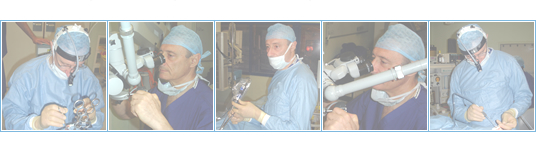
Common surgical procedures
Removal of the tonsils, or adenotonsillectomy is indicated in the following situations:
- Recurrent tonsillitis (more than 4 attacks per year);
- Obstructive sleep apnoea;
- Tonsillar enlargement;
- Hypertrophy of tonsils and adenoids in children;
- Peritonsillar abscess with a past history of recurrent tonsilliltis;
- Persistent sore throat after glandular fever;
- Unilateral enlarged tonsil with lymphoma as a possibility.
Although this is one of the most common performed operations it is not without complications. Haemorrhage is the most important may be primary within 24 hours of surgery or secondary (between 5 and 10 days) when it is usually the result of infection in the site of excision. Bleeding disorders such as von Willebrand's disease may present with bleeding during or after tonsillectomy.
|